A quick approach to Pneumothorax
Topics / Surgery / Trauma / Pneumothorax
Definition
The accumulation of air inside the pleural space (between the visceral and the parietal pleura).
Basics
Air enters into the pleural space due to injury of the airway or the chest wall. The injury results in the loss of intrathoracic negative pressure, the lungs are no longer kept expanded. The injury may form a one way valve mechanism that allows air to enter into the pleural space, but it prevents it from escaping. As time progresses, this accumulated air increases in volume and it will progressively decrease the volume of the lung. If the disease process continues, the increased air pressure causes compression of the intrathoraxic structures (obstructive shock). There is reduced venous return and reduced cardiac output.
Types
- Traumatic pneumothorax
- Size
- Small Pneumothorax ( < 2cm)
- Large Pneumothorax ( > 2cm)
- It must be said that there are many different ways to measure the size of a pneumothorax. Some sources may suggest the apex-to-cupola distance (in this case use 3cm instead of 2cm), interpleural distance at the level of the hilus of the lung or Collins method (%).
- Size
- Mechanism of injury
- Blunt chest trauma
- Penetrating chest trauma
- Iatrogenic (during mechanical ventilation, CVP insertion, lung biopsies)
- Type
- Close pneumothorax: The air will enter the pleural space via the airway, usually due to blunt trauma.
- Open pneumothorax: There is a connection between the environment and the pleural space due to an injury in the chest wall.
- Non-Traumatic (Spontaneous Pneumothorax)
- Primary (The patient has no underlying disease)
- This is due to the rupture of subpleural blebs, the risk is higher in certain groups of individuals such as those who are smokers.
- Secondary (The patient has an underlying disease)
- Common causes are rupture of bullae (emphysema) in COPD, Infections (Pulmonary TB, PJP), cystic fibrosis, malignancy
- Primary (The patient has no underlying disease)
Presentation
- Asymptomatic
- Acute onset of shortness of breath
- Ipsilateral pleuritic chest pain, visible injury
- Subcutaneous emphysema
- Hyperresonant ipsilateral side
- Decreased breath sounds on the affected size
- If tension pneumothorax (Distended neck veins, Trachea deviates to the opposite side, signs of obstructive shock – tachycardia, hypotension, pulsus paradoxus)
Diagnosis
- Tension pneumothorax is a clinical diagnosis, then perform an immediate chest decompression.
- Chest X-Ray (preferably Erect PA on inspiration, supine has decreased sensitivity)
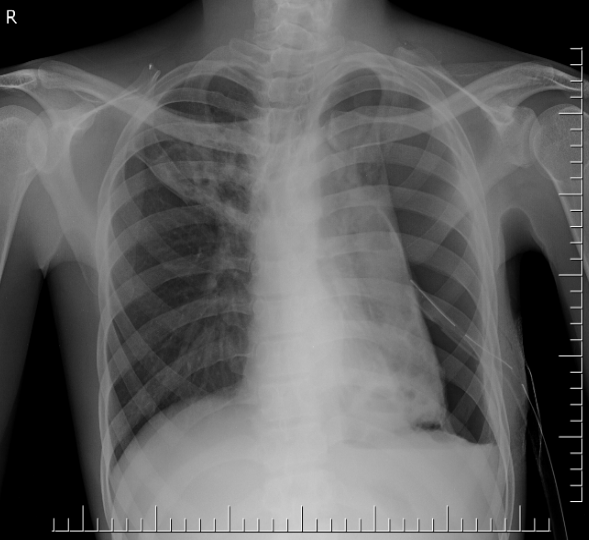
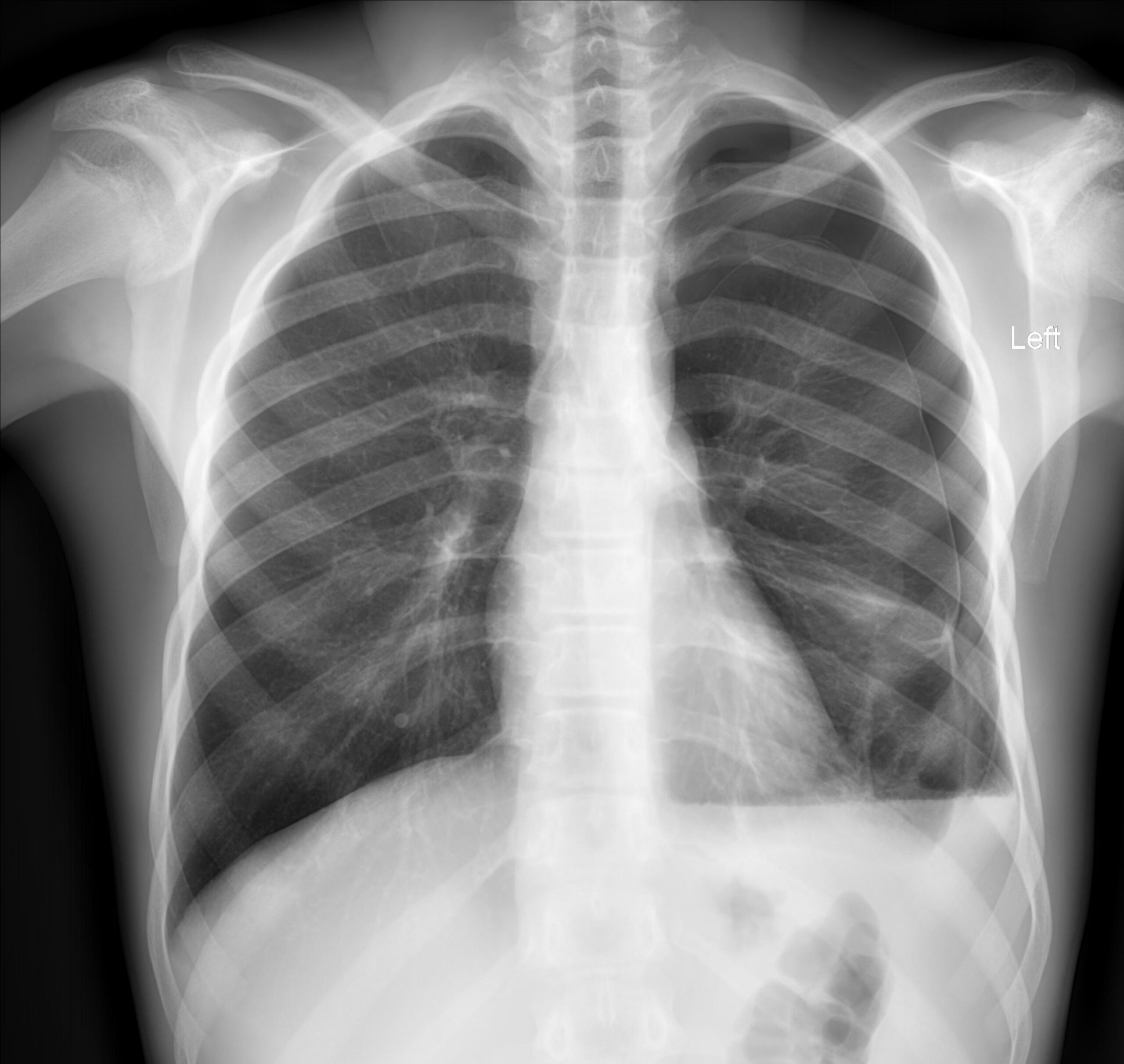
- Usually there is visible ipsilateral visceral pleura line and reduced lung markings with changes in radiolucency. If there is tension pneumothorax you may see ipsilateral flattening of the diaphragm, widening of the intercostal spaces, mediastinal shift to the contralateral side, deviation of the trachea to the contralateral side), deep sulcus sign
- Ultrasound (as part of eFAST)
- Air accumulates between the visceral and the parietal pleura. This means that when an ultrasound probe is placed on the chest well there will be (1) abscence of the pleura sliding since there is now a space filled with air (2) prominent A-lines (horizontal lines) (3)absence of B-lines (vertical lines) (4) barcode sign instead of the seashore sign in the M-mode
- CT Chest – especially for the non-traumatic patients, this will provide some information for the cause.
Management
1. Small Pneumothorax
Usually < 2cm in size, measured from the inside of the third rib to the margin of the lung. These patients should be treated without an intercostal drain if the following is true:
- The small pneumothorax is unilateral
- The patient is not ventilated/intubated
- The patient does not require any anesthesia
You will then need to review the patient and repeat a chest X-Ray after around 3-6 hours. If the pneumothorax has not increased in size then the patient may be discharged. This is because a small pneumothorax will resolve by itself, especially in around 10 days. However, if the size of the pneumothorax has increased in size then you should insert an intercostal drain. However, if the patient has a small pneumothorax and is unstable, the patient may benefit from an ICD. The ICD is inserted in the 5th intercostal space (bounded by the 5th and 6th rib, insert the ICD along the superior border of the 6th rib [the rib below] to avoid the neurovascular structures), along the anterior axillary line (to avoid the long thoracic nerve) on the ipsilateral side.
2. Large Pneumothorax
Usually, the size is > 2cm in size, this one requires the insertion of an ICD, which is described below.
3. Tension Pneumothorax
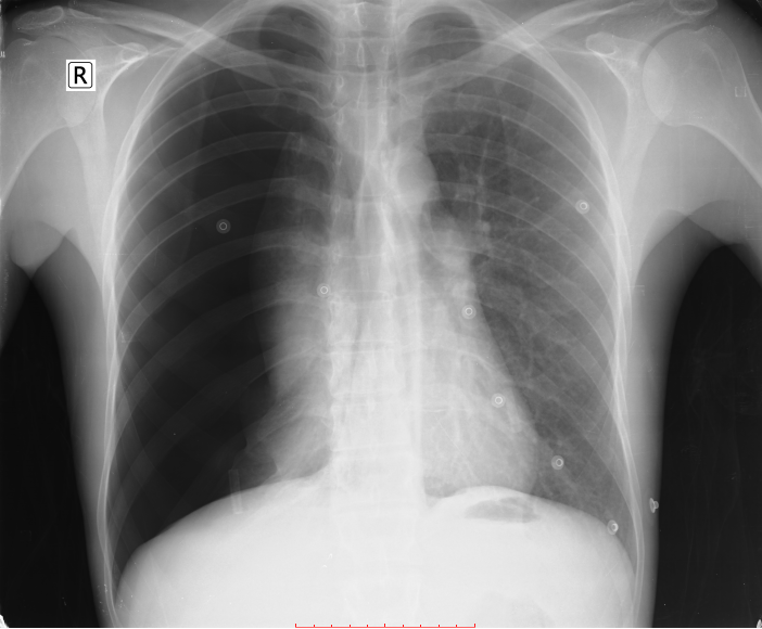
This pneumothorax has become so severe that there is increased tension, compressing the other structures inside the chest, especially the mediastinal structures. This is a clinical diagnosis and the patient will present with the following:
- Respiratory signs: Severe respiratory distress (Nasal Flaring, tachypnea, use of accessory muscles of respirations, desaturation on room air), trachea deviates to the opposite side. Cyanosis is a late sign. There will be hype-resonance on the side of the tension pneumothorax and decreased air entry on the side of auscultation
- Signs of shock: Tachycardia, hypotension
Step-wise Management
- ATLS principles: ABCDE
- Immediate chest decompression with by performing a needle thoracostomy (Insert a large bore IV cannula into the second intercostal space, midclavicular line OR 4th-5th ICS in anterior axillary line.) This is a temporary form of management, the definitive treatment is the insertion of an intercostal drain (ICD) which occurs afterwards. The ICD should be a large bore, adult size around 36 – 40 Fr.
- Then, review results and admit the patient
- Position the patient in a upright position since this improves respiratory function
- Supplemental oxygen to keep saturations of oxygen > 92%
- Mobilize as soon as possible
- Chest physiotherapy and breathing exercises. You can ask the patient to blow into a glove and you may also consult physiotherapy.
- Removed the ICD once the lung has expanded. You should assess this by repeating an chest X-Ray and also correlate it clinically. The ICD drain has an underwater seal which should have stopped bubbling (this is an indication that it may be removed). There are other types of ICDs that do not display this mechanism and will have other indicators.
Depending on the assessment other departments may need to be consulted: Pulmonology, Cardiothoracic surgery, ICU
4. Open Pneumothorax
- Apply a partially occlusive dressing over the lesion. You can make this by occluding 3 sides of a 4-sided dressing. This will create a one-way valve mechanism that allows air to escape the chest but it will not allow the air to enter the pleural space.
- Insert an ICD (Tube thoracostomy).


Made with ❤️ by QuickBeat
We are a small education agency that aims to provide medical education.
EXPLORE
SERVICES
CONTACT
- Pretoria North, Gauteng
- contact@quickbeat.co.za