Diabetic Foot Sepsis

Definition

Diabetic foot sepsis can be defined as an infected foot that has been affected by peripheral neuropathy and/or peripheral arterial disease of the lower limb in a patient with diabetes. The severity of the infection may vary from cellulitis to gangrene.

Basics

The main mechanisms involved in the development of diabetic foot sepsis include:

  1. Peripheral Neuropathy
    • Autonomic Neuropathy: There is a lower level of perfusion because of a lack of adequate capillary recruitment and there is also increased shunting of blood around the capillary bed. The sweat and oil glands are also affected and this results in dry skin that is prone to “cracking”.
    • Sensory Neuropathy: Loss of peripheral sensation prevents the patient from responding adequately to a nociceptive stimulus. This means that minor injuries usually go unnoticed by the patient until they are severe.
    • Motor Neuropathy: There is associated muscle weakness and foot deformity that affects the joints and pressure points. Glycosylation of tendons causes stiffening and shortening of several tendons which result in various deformities (claw toes, hammer toes). These deformities also cause changes in pressure points.
  2. Vascular disease
    • Macrovascular disease: Diabetic patients have accelerated atherosclerosis resulting in poor circulation and decreased wound healing.
    • Microvascular disease: There are mainly two hypotheses: The “hemodynamic hypothesis” and the “capillary steal syndrome”. The hemodynamic hypothesis suggests that blood flow dysregulation results in structural alterations such as thickening of the basement membrane and microvascular sclerosis. In the capillary steal syndrome, as explained above, autonomic neuropathy affects the sympathetic innervation to the microvasculature and results in the shunting of blood away from the capillaries.
  3. Infection
    • Usually, start as an infection caused by Staphylococcus or Streptococcus but as the infection progresses it quickly becomes a polymicrobial infection. In the last two stages, there is a significant increase in gram-negative and anaerobic microorganisms. Diabetes impairs the immune system to fight any infection. It causes a decrease in leukocyte migration (polymorph), decreases in phagocytosis, decreases in intracellular killing, and a decrease in chemotaxis. Treating an infected diabetic foot with antibiotics is very difficult. This is because in many cases, the vascular supply to the area has been affected and it reduces the ability of the antibiotic to read the target site.

The Polyol Metabolic Pathway

Risk Factors

The following is a list of factors that increases the overall risk that the patient will develop Diabetic Foot Sepsis.

  1. Charcot joint/Charcot arthropathy
  2. Peripheral vascular disease
  3. Microvascular Complications: Peripheral Neuropathy, Retinopathy or Nephropathy
  4. Macrovascular Complications: History of cerebral vascular disease, Coronary Heart Disease
  5. Diabetes Mellitus > 10 years
  6. The use of insulin (means the diabetes is more severe or advanced), Poor glycemic control
  7. Age > 45, Male Gender
  8. Smoking, Hypertension, Overweight, Obesity

Presentation

The patient may present with symptoms and sign suggestive of having neuropathy, vascular disease, infection (local or systemic), tissue loss, or a deformity. The patients may also present as an emergency in the form of sepsis or septic shock or from complications of Diabetes Mellitus such as DKA/HHS. Having performed the necessary questions and assessment, you should be able to grade the patient using Wagner’s Classification. Always remember that Diabetes is a disease that affects many organs and a thorough history and examination is required. Read how to approach an ulcer in order to learn more about what questions to ask and how to examine this patient.

Classification

Meggitt–Wagner’s Classification
Ulcer Grading Description
Grade 0 No ulcer. Intact skin. But the foot is at high risk. Not infected. Any post-ulcerative lesion that has been completely epithelialized.
Grade 1 Superficial, full thickness ulcer which is limited to the dermis, not extending to the subcutaneous tissue. Not infected.
Grade 2 Deep ulcer, extending through the subcutaneous tissue with exposed tendons, ligaments, joint capsules, deep fascia or bone but no osteomyelitis or abscess formation. Cellulitis is included here.
Grade 3 Abscess, joint sepsis or bone involvement (osteomyelitis)
Grade 4 Localized gangrene of the toes or forefoot
Grade 5 Extensive Gangrene involving the whole foot

Investigations

  1. Blood
    • Formal Blood: Full Blood Count, Septic Screen, Urea and Electrolytes with Creatinine, HbA1C (assess glycaemic control over the last 3 months)
    • Blood Gas: Metabolic acidosis, high sugar level, high lactate
  2. Cultures
    • Take a deep tissue culture as superficial colonization of the wound may not be representative. Send this wound swab or tissue biopsy for microscopy, culture, and sensitivity.
  3. ECG
  4. Imaging
    • X-Rays of the affected limb: This is to rule out osteomyelitis, but one should remember that x-ray changes for osteomyelitis take around 4-6 weeks to develop.
    • Dopper/Duplex Ultrasound to assess for flow.
    • Angiography
    • CT Scan of the affected limb: May assist with the identification of deep space abscesses or collections.
    • MRI Scan of the affected limb: This may assist in the detection of osteomyelitis earlier if there is any suspicion.
  5. Other:
    • Transcutaneous Oxygen Measurement (TCOM): This is sometimes also known as “Transcutaneous Partial Pressure of Oxygen”. It is a non-invasive way of measuring the quantity of skin oxygenation. This is a measurement of the oxygen tension from around 1 to 2mm deep in the local skin capillaries. We place the probe next to an ulcer and this will help us predict wound healing and the patient’s possible response to hyperbaric oxygen therapy. For instance, the normal oxygen tension ranges from 50-70mmHg. An oxygen tension < 40mmHg will cause impairment of wound healing. Oxygen tension < 30mmHg classifies the patient as having Critical Limb Ischemia.
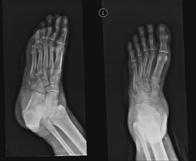
"Diabetic foot and secondary osteomyelitis. Destruction of the base of 5th metatarsal with moth eaten appearance and loose debris. Non visualization of the 3rd and 4th tarsometatarsal joint space. Loss of normal tarsometatarsal joint planes. The intermediate and lateral cuneiform, and the cuboid intertarsal planes are lost." Case courtesy of Dr Varun Babu, Radiopaedia.org. From the case rID: 46417
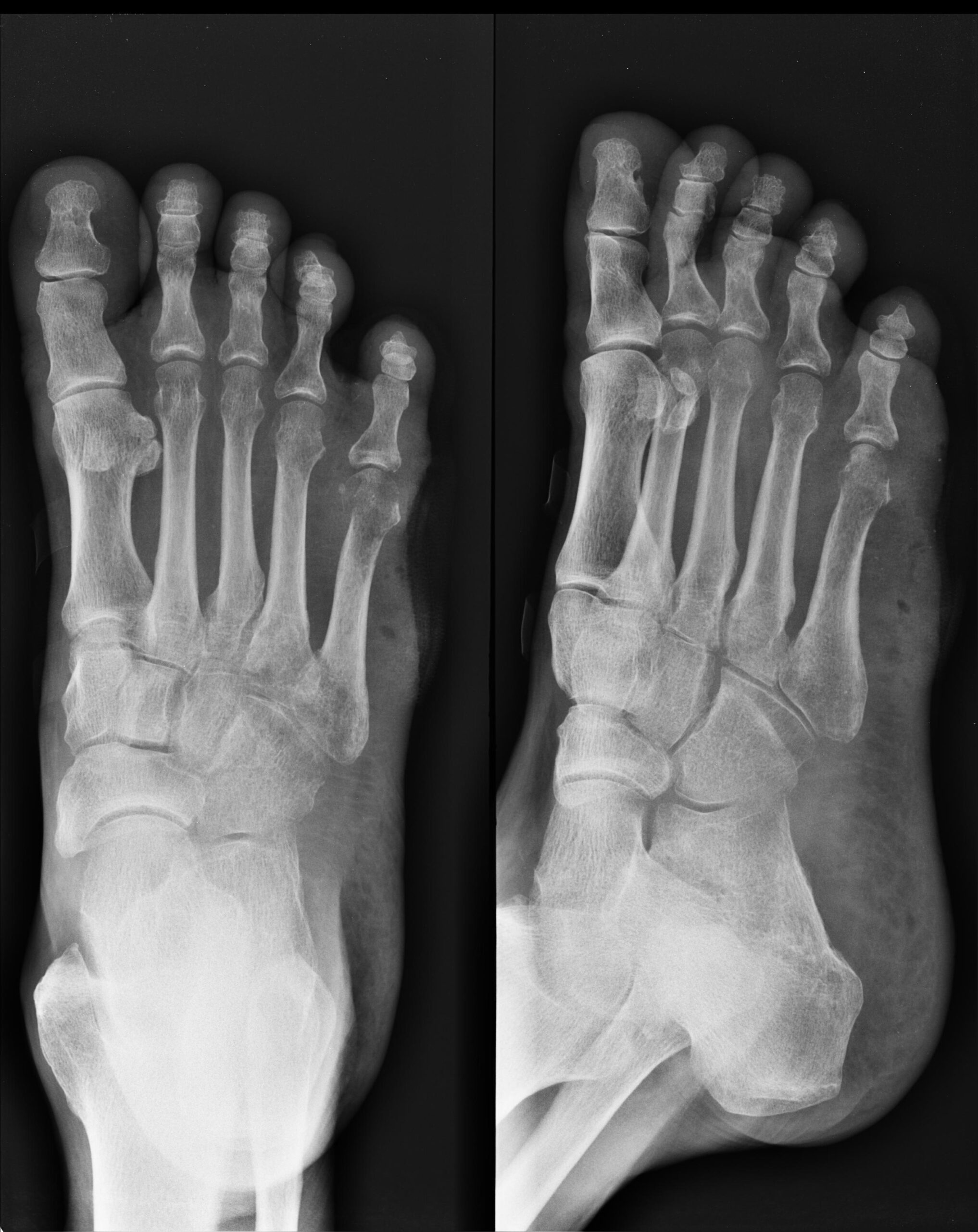
"Periarticular osteopenia involving 2nd to 5th MTP joints. Lytic area in 5th metatarsal head - neck with cortical breach. Soft tissue swelling with air foci in mid foot laterally. NO arterial calcification seen."Case courtesy of Dr Maulik S Patel, Radiopaedia.org. From the case rID: 18692

Management

All diabetic patients should have a specialized diabetic foot examination and ideally should also be seen by a podiatrist. These patients should be managed together with a multidisciplinary team. The surgical management of Diabetic Foot Sepsis depends on the Wagner Classification. It should be noted that many patients develop Diabetic Foot Sepsis on the contra-lateral limb within 20 months of amputation. This means that these patients should be extensively managed by the multidisciplinary team even after an amputation to prevent this from happening.

  • Grade 0:
    • This foot requires treatment with prevention and prophylaxis
    • Educate the patient
    • Counsel on footwear
    • Regular diabetic foot examination
  • Grade 1:
    • Antibiotics
    • Control of co-morbidities (eg glycaemic control)
    • Pressure Relief: Accommodative footwear, total contact cast/splint, walking brace, crutches, wheelchairs
  • Grade 2:
    • Surgical Debridement
    • Wound dressing
    • Antibiotics
    • Control of co-morbidities (eg glycaemic control)
    • Pressure Relief: once the wound heals to Grade 1
  • Grade 3:
    • Surgical Debridement with some form of amputation may be required in some cases.
    • Wound dressing
    • Antibiotics
    • Control of co-morbidities (eg glycaemic control)
    • Pressure Relief: once the wound heals to Grade 1
  • Grade 4:
    • Extensive debridement and amputation (two-stage amputation). The patient may benefit from revascularization procedures.
    • Wound dressing
    • Antibiotics
    • Control of co-morbidities (eg glycaemic control)
  • Grade 5:
    • Below knee amputation
    • Wound dressing
    • Antibiotics
    • Control of co-morbidities (eg glycaemic control)

Prevention

All patients should be educated about good lifestyle modifications guided by the Diabetes Management Guidelines. This includes but is not limited to the cessation of smoking, cessation of ethanol use, encouraging weight loss, exercise, dietary modification, diabetic foot examination, protective foot wear, and diabetic foot care.

Complications

  1. Infections: Cellulitis, Gangrene, Osteomyelitis
  2. Ulceration, Loss of tissue
  3. Amputation
  4. Deformity
  5. Disability
  6. Death
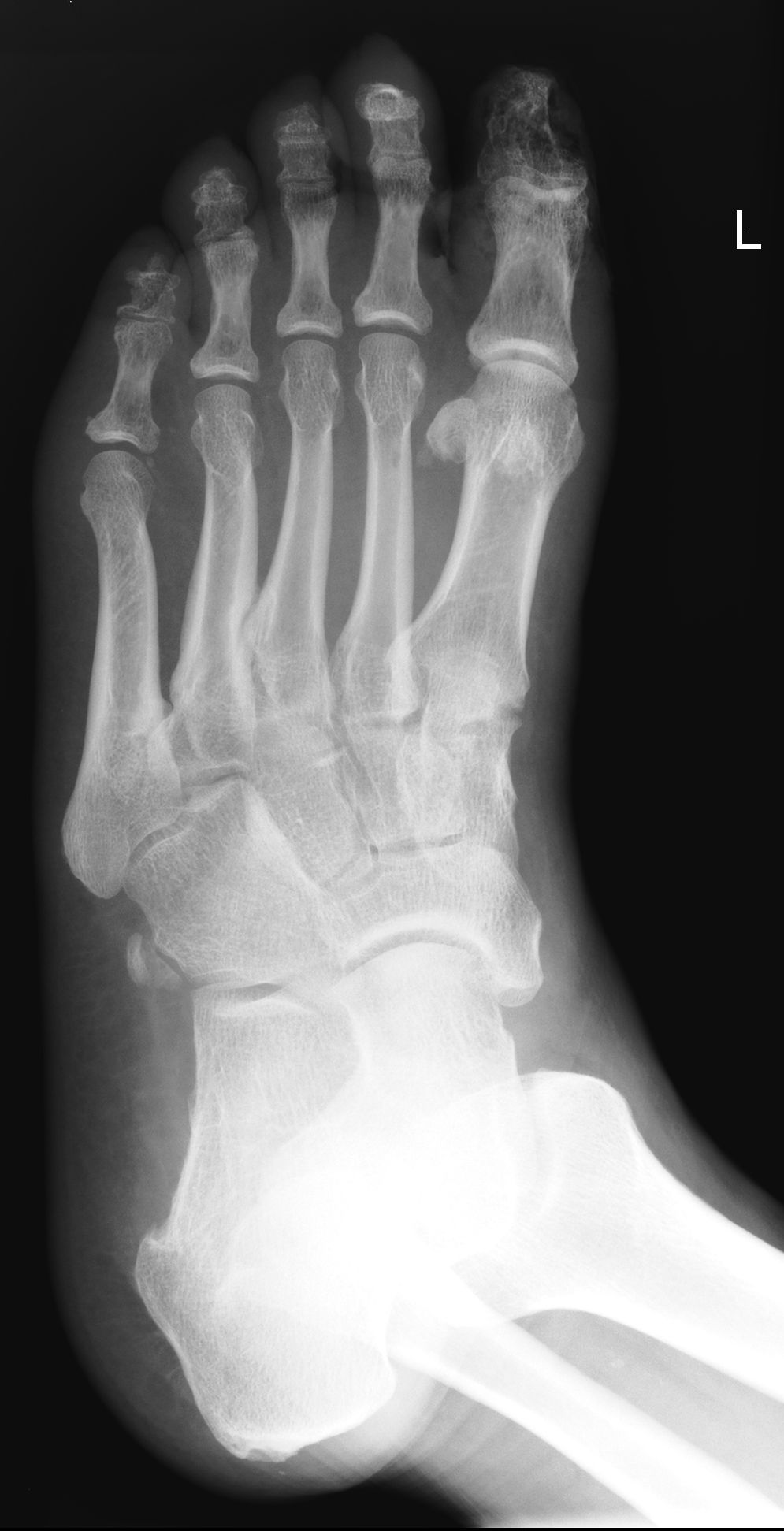
""There is loss of soft tissue over the great toe, with further lucencies in the surrounding soft tissue. This is very suggestive of superimposed infection. The underlying phalanx shows patchy osteoporosis and is suggestive of osteomyelitis." Case courtesy of Assoc Prof Frank Gaillard, Radiopaedia.org. From the case rID: 7663
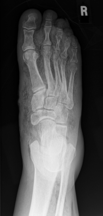
"Necrotizing Fasciitis in a Diabetic Foot. There is extensive subcutaneous emphysema tracking along the facial planes throughout the foot and distal leg. Note that the proximal extent of the subcutaneous gas is not visualized." Case courtesy of Dr Matt Skalski, Radiopaedia.org. From the case rID: 25026

Important Image

Some Text

Articles

Made with ❤️ by QuickBeat

We are a small education agency that aims to provide medical education. 

CONTACT

Copyright © 2022 Quickbeat​. All Rights Reserved.