Intussusception
Defintion
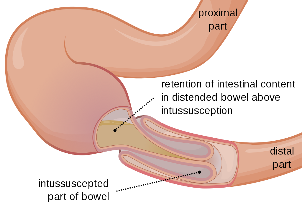
Intessusception occurs when there is an invagination of the proximal bowel (intessuceptum) into the distal bowel (intussuscipiens). Yeah, so it is basically like one of those “old telescopes”. The proximal segment goes inside the distal segment.
Basics
The proximal segment moves into the distal segment by peristaltic activity. There is usually a lesion that increases the chances of this from occurring, this is called the “lead point”. The lead point is often trapped by the peristaltic movements and dragged into the distal segment of the intestine resulting in intussusception. However, it is worth noting at that not all forms of intussusception will have a lead point.
As the proximal segment is brought into the distal segment, the mesentery that is pulled inside will become compromised. This results in venous obstruction and in bowel wall oedema. This is then followed by arterial insufficiency and it will lead to ischemia and bowel necrosis. Therefore, this highlights the importance of treating this condition quickly.
Intussusception is uncommon in patients that are below 3 months old and above 3 years of age. The highest incidence occurs in children with the ages 4 – 9 months old.
Classification
- Primary Intussusception:
- This type of intussusception is caused by hypertrophy of the Peyer’s patches. Since the Peyer’s patches are made of lymphatic tissue, it makes sense that this usually occurs after a recent history of an upper respiratory tract infection or gastroenteritis.
- Viruses, such as Adenoviruses and Rotaviruses have been also responsible for the hypertrophy of the Peyer’s patches.
- Previous immunization with the Rotavirus vaccine is also a risk factor for the development of this type of intessusception.
- Secondary Intussusception:
- This occurs when the “lead point” is identified.
- Common lead points:
- Benign: Meckel diverticulum, polyps, the appendix, hemangiomas, carcinoid tumors, foreign bodies and so forth.
- Malignant: Rare, may include lymphomas and small bowel tumours.
Presenation
- Intermitted, crampy abdominal pains associated with a “currant jelly” stools. The “currant jelly” stools is usually a late sign. An earlier sign may be the appearance of blood tinged stools. Therefore, a per rectal examination is important.
- The child will get intermittent episodes of pain, and in-between these episodes the child appears to be fine.
- Palpable abdominal mass (sausage shape or curved) on physical examination.
- The right lower quadrant may appear to be empty or flat as the intussuscepted mass moves in a cephalic direction. This is called the Dance sign.
- As the intessuceptum progresses, it may result in prolapsed of the intessuceptum through the anus. (Often misdiagnosed as a rectal prolapse). If a tongue blade can be inserted more than 2cm into the anus along the sides of the prolapsed mass then one can make the diagnosis of intussuception.
- Other symptoms: bowel obstruction (bilious vomiting, abdominal distension, no passage of stools).
Investigations
- Bloods
- Formal bloods: Urea, Electrolytes and Creatinine.
- Blood gas
- Imaging:
- Abdominal X-Rays (erect) – may find an abnormal distributuion of bowel gas, air-fluid levels, abdominal mass. However, x-rays cannot be used alone to do a diagnosis.
- Ultrasound: This is used a screening tool. Findings may include a “target” or “
Investigations
Lorem ipsum dolor sit amet, consectetur adipiscing elit. Ut elit tellus, luctus nec ullamcorper mattis, pulvinar dapibus leo.


Made with ❤️ by QuickBeat
We are a small education agency that aims to provide medical education.
EXPLORE
SERVICES
CONTACT
- Pretoria North, Gauteng
- contact@quickbeat.co.za