An Approach to Breast Cancer
Basics
Anatomy
1. Medial mammary branches of perforating
branches and anterior intercostal branches of the internal thoracic artery (arises from subclavian).
2. Lateral thoracic and thoracoacromial arteries (arises from axillary artery).
3. Posterior intercostal arteries (arises from the thoracic aorta in the 2nd, 3rd and 4th intercostal
space).
1. Axillary vein
2. Internal thoracic vein
1. Axillary Lymph nodes – 75% of the ipsilateral breast drains to the axillary lymph nodes. There
are 5 groups: Anterior, posterior, medial, lateral and apical lymph node group. These lymph nodes
will then drain towards the supraclavicular, infraclavicular and cervical lymph nodes.
- The lymph nodes are anatomically divided into levels by the pectoralis minor muscle:
- Level 1: Lateral to the pectoralis minor
- Level 2: Posterior to the pectoralis minor.
- Level 3: Medial to the pectoralis minor, extending up to the apex of the axilla.
2. Internal Mammary nodes – Drain 20% of the breast, especially the upper and lower inner
quadrants.
3. Interpectoral Lymph nodes – Located between the pectoralis major and the pectoralis minor
muscle
Differentials (Breast Lump)
The differentials for a breast lump can be classified according to the pain. It is important to note that breast malignancies are present more commonly as painless breast lumps.
| Painless Lump | Painful Lump |
|---|---|
|
|
Pathology of Breast Cancer
WHO classifies breast cancers into epithelial and non-epithelial tumours. Non-epithelial tumours arise from the supporting stroma of the breast and these include angiosarcoma, malignant phyllodes tumour,s and primary sarcomas. The epithelial tumours arise from the cells lining the ducts or lobules. They can be divided into invasive and non-invasive based on the invasion of the basement membrane.
| Differential for Breast Cancer | ||
| Non-Invasive | Invasive | |
|---|---|---|
| Ductal | Ductal Carcinoma in Situ (DCIS) | Invasive Ductal Carcinoma (IDC) |
| Lobular | Lobular Carcinoma in Situ (LCIS) | Invasive Lobular Carcinoma (ILC) |
| Others | 1. Medullary, colloid,
tubular, papillary 2. Inflamatory carcinoma of the breast (T4d). |
|
I have omitted details concerning each of these cancers on purpose. These notes will be focusing on the clinical aspect of breast cancer and not necessarily on the in-depth details as you would have been expected to know in year III of your MBChB curriculum.
History
1. History of Presenting Complain
2. History of Risk Factors
3. History of Metastatic Work Up
4. History of Patient Operability
The presentation of the patient
- Erythema
- Peau d’ Orange – “Orange peel skin”. Caused by
lymphatic obstruction. - Ulcers
- Skin colour changes – Could be caused by poor
lymphatic drainage or inflammation. - Warmth of skin, Swelling.
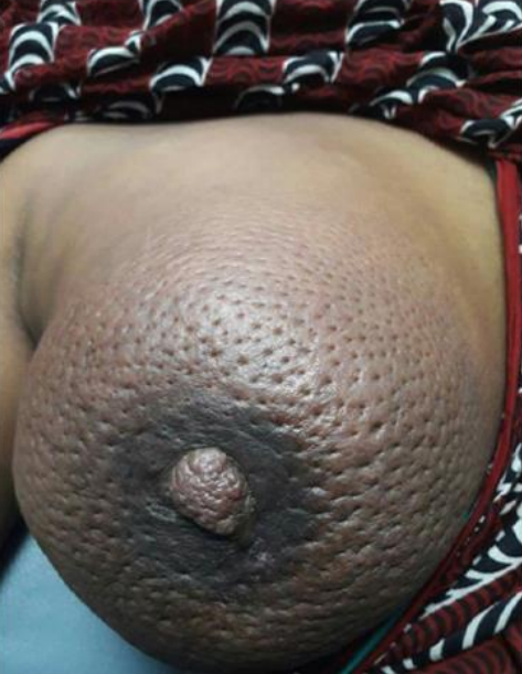
The patient may also complain of breast pain without a lump. Breast pain could be cyclical or noncyclical. Cyclical means that it follows the menstrual cycle and it is common among women. If it is non-cyclical then it usually means that there is some pathology. Breast pain is termed mastalgia or mastodynia. Mastodonia also refers to breast pain, but it may refer to breast pain in males (Mtimba L, 2018). However, the term mastalgia is the one commonly used. Non-cyclical pain is usually due to malignancy until proven otherwise.
Tethering, also known as skin dimpling may also be present. There is a dimple over the lump and this occurs because as the tumour grows, it encroaches on the connective tissue and “pulls” the suspensory ligaments of Cooper to make the appearance of a dimple over the skin. Although it is seen on the skin, it is actually NOT part of skin changes.
The most important presentation is saved for last. The patient can also be symptomatic. This presentation is common in “large – obese” patients.
The history of the Presenting Complain
You need to ask about the different types of presentations mentioned above. These questions are
simple to remember if you understand how they can present.
- Painless breast lump?
- History of nipple discharge?
- If present – ask about the colour:
- Clear – Cystic
- Yellow/Pus – Abscess/ Mastitis
- Red or pink – Ductal papilloma/carcinoma
iv. White fluid – Galactorrhoea/Lactation - Remember that during your investigations, you may want to send a sample for
cytology/MCS. If the discharge is bilaterally it may refer to hormonal imbalances.
- If present – ask about the colour:
- Any skin changes?
- Any pain? – Remember to define if it is cyclical or non-cyclical.
- Areolar and nipple issues?
- You may ask about metastatic complains here as well.
- Back pain?
- Difficulty in breathing?
- Fits
- Headaches
- But this has its own section, so you don’t need to. The reason I have included it here is that in a patient with advanced breast cancer these symptoms may form part of the main presenting complaint.
For each of these presentations, ask about the onset, location, duration, progressiveness, is it intermittent or persistent, unilateral or bilateral, and so on.
History of Risk Factors
The risk factors could be hormonal or non-hormonal. You need to find out if your patient has any of these
1. Non-Hormonal History
- Being a female has an increased risk due to their breast tissue and general female behavior.
- Age – The older you become the higher the risk. However, malignancy in the young tends to
be more aggressive than when you’re old. - Previous history of cancer:
- A personal history of cancer?
- A family history of cancer?
- This is important especially if it is a first degree relative. BRCA 1 and BRCA 2 cancers are very important: Ovarian, Colon, Skin, Endometrial, and so on. Maternal side cancers are very important. The age of the family member affected by breast Cancer is also important because:
- If young – hereditary (There’s a pattern in the family tree).
- If old – sporadic.
- How to ask the questions:
- Do you have a history of breast cancer in the family?
- Which one was it?
- How old was the person in your family when they had it?
- This is important especially if it is a first degree relative. BRCA 1 and BRCA 2 cancers are very important: Ovarian, Colon, Skin, Endometrial, and so on. Maternal side cancers are very important. The age of the family member affected by breast Cancer is also important because:
- Radiation exposure – Had you had any history of radiotherapy?
- Obesity (Can also be hormonal)
- Alcohol
- Smoking
- Breast lesions – Fibroadenomas increase the risk.
- Race – White people have an increased risk but are generally detected earlier than in black
people. Black people have a lower risk but the malignancy is detected very late and in
advanced stages. - Breast tissue density – In old women, the breast tissue density is lower than in young
women.
2. Hormonal History
Hormonal risk factors can be grouped under:
- Gynecological History
- Past-Obstetric History
- Miscellaneous
Gynaecological History
- Early menarche (<12 years old) with late menopause (>55) is a risk factor. This is called the “Estrogen window”, what is important to know is that estrogen is a carcinogen. A prolonged exposure to it will increase the risk of malignancy.
- Tips: A good way to ask for menopause after pregnancy has been ruled out or any other diseases: “When was your last menstrual period?”
- Using contraceptives – ↑ Hormones
- Hormone replacement therapy – These hormones should ideally not be given to a patient before consulting with a breast surgeon so that a risk assessment may be made.
- Other exogenous sources of hormones you can think of.
Past-Obstetric History
- Parity – nulliparity increases the risk.
- Breast feeding – Breast feeding will decrease the risk.
- First pregnancy – pregnancy in teenagers will decrease the risk.
- If age of first pregnancy >25-30: ↑ risk.
- If age of first pregnancy >30-35: ↑↑ risk.
- Pregnancy in advanced ages > 35-40: ↑↑↑ risk.
- Termination of Pregnancy(TOPS)
- When the breast grows during pregnancy, it occurs similar to how breast tumours
grow during breast cancer. It grows due to hormonal exposure. If the pregnancy is
terminated suddenly, you do not give a chance for breast development to terminate
accordingly and hence this may increase the risk of malignancy.
- When the breast grows during pregnancy, it occurs similar to how breast tumours
Miscellaneous
- Obesity – The fatty tissue is able to convert androgens into estrogen by aromatization.
- Fatty food.
Metastatic History
- Weight loss and loss of appetite – Common in any malignancy.
- Bone pain?
- The most common bone pain arises in the back. This is due to vertebral
metastases. As the tumour grows it may also compresses the nerves and leads to
neurological issues below the level of compression.
- The most common bone pain arises in the back. This is due to vertebral
- Chest
- Difficulty in breathing?
- Coughing – productive or non-productive? Breast malignancy can metastasize to the chest and cause:
- Ipsilateral pleural effusions
- Cannonball opacities
- Hilar lymphadenopathy
- Bone lesions
- Abdomen – a common site for metastasis is the liver. Which may result in an abnormal
liver function. You will need to ask questions related to liver function:- Tenderness?
- Discoloration of the skin, and mucous membranes? – Trying to rule out jaundice.
- Distension of the abdomen? – Trying to rule out ascites.
- Brain – You will need to ask questions related to the CNS.
- Headache?
- Blurred vision?
- Abnormal gait? Cancer can lead to leptomeningeal carcinomatosis (LC), a rare complication of
cancer in which the disease spreads to the meninges surrounding the brain and
spinal cord.
- Skin – Skin satellites.
- Endocrine issues – Also possible, however, the above-mentioned are the most important.
History of Operability
- Active person?
- Exercise? – Please understand that exercise is not just about going to the gym and lifting
weights. Walking, carrying buckets of water, gardening and other activities may be
assessed as “exercise” for the people of the community. - Co-morbidities?
- Cough?
- Cardiac issues?
- Previous chest injuries?
- Difficulty in breathing lying down? How many pillows do you need?
- Exclude current and previous chest issues – TB? If a person had TB before, their lungs will not be functioning as well as a healthy person.
Staging
| TNM Classification | ||
| Tumour Size | Nodal Involment | Metastasis |
|---|---|---|
| Tx:Tumour could not be assesed – occurs when we cannot measure the tumour or are only able to stage it radiologically instead of clinically. | Nx: Nodes could not be assessed – This could be due to extrammary tissue present in the axillary tail of Spence. When the patient goes into her menses/breastfeeds the axilla becomes engourged. This prevents adequare palpation of the axilla. This can also be due to infections/skin conditions that prevent exammination. | M0: No metastasis present. |
| Tis: Carcinoma in situ. | N0: No lymph node involment. | M1: Metastasis is present. This could include: 1. Bone – especially vertebrae 2. Chest –lungs 3. Abdomen – liver 4. Skin 5. Brain |
| T1 – less than 2cm: a: 0.1 – 0.5cm b: 0.5 – 1cm c: 1 – 2cm |
N1: Ipsillateral mobile axillary lymph node involment. | |
| T2: 2 – 5cm | N2: Ipsillateral fixed/matted axillary lymph node involment. | |
| T3: more than 5cm | N3: Ipsillateral supraclavicular/infraclavicular lymph node involment or contrallaterl or internal mammary lymph node involment. | |
|
T4: Any size with chest or skin
involment, or both. a: Chest wall involment – This will include pectoralis major, serratus anterior, intercostal muscles and ribs. If it only includes the pectoralis major then it is not a T4a lesion but rather T3. b: Skin involment – ulceration, discolorations, ulcerations, peau orange and areolar and nipple changes. c: Chest and skin are both involved. d: Inflammatory breast cancer – The breast will have the typical signs of inflammation such as rubor, dolor, calor and tumor. A differential for this would be mastitis. |
||
Physical Examination in Summary
Breast Examination
Inspection
Ask the patient to put both arms on their sides and inspect both breasts. The patient may be sitting or standing. Assess for symmetry, masses, size of the breasts, skin changes, alveolar and nipple changes, and dimpling. The patient should then be asked to lift their arms 90 to 180 degrees to assess any changes in this position.
Palpate
Palpate all the lymph nodes first. Palpate the lymph nodes of the head, neck, supra/infraclavicular, and axillary lymph nodes.
Read this part properly:
- To examine the patient’s right axilla: Handshake your patient’s right hand with your right hand.
Use your left hand to palpate the patient’s right axilla while letting their elbows resting on your left
forearm. - To examine the patient’s left axilla: Handshake your patient’s left hand with your left hand. Use
your right hand to palpate the patient’s left axilla while letting their elbows rest on your right
forearm.
After the palpation of the lymph nodes, put the patient in a supine position. Go to the side of the pathologic breast and ask them to put their hand behind the head. This will stretch the muscles allowing you to palpate more accurately. Palpate each quadrant of the breast for any breast lumps and notice their size, location, mobility, and consistency. You may also palpate for tenderness of the breast.
Lastly, assess the nipple by asking the patient if they could squeeze them in order to observe any discharge. Remember to repeat the breast examination on the contralateral side.
Systemic Examination
In this section, you will be examining the patient in order to rule out any metastasis on a clinical basis. You will need to examine the patient for any bone pains as the malignancy of the breast commonly metastasizes to the vertebrae. Then you will need to focus on the examination of the chest (respiratory system examination), the abdomen, the central nervous system, and the skin.
At the end of the clinical examination, you should be able to say, “This is clinically a TaNbMc” or “This is a Stage 1/2/3/4 breast cancer”.
Investigations
The reasons we do investigations:
- To Diagnose
- To Stage
- To Assess the Operability of the patient
A breast Cancer diagnosis is done using the triple assessment. This includes:
- Clinical Examination (History and Physical Exam)
- Imaging
- Histology (Tissue Diagnosis)
A.Imaging Modalities
- Ultrasound
- Mammography
- Breast MRI
An Ultrasound will be used for every woman, however, in women that are older than 40 years of age, mammography is added to the imaging modalities used. Women that are younger than 40 will only be investigated with Ultrasound imaging. The women that are young, pregnant, or lactating have a breast tissue density that is high. If you do mammography on these women the results of the mammography will come as very “white”, obscuring the lump that also looks “white”. Hence for these women we only use Ultrasound. Older women have a decreased breast tissue density and hence mammography can be done. In summary, we do ultrasounds on young women. We do Ultrasound and Mammography in older women.
| Ultrasound | Mammography | Breast MRI |
|---|---|---|
2. Localization of the mass 3. Assess the mass in areas that mammogram cannot access, such as the axilla. 2. With hyperechoic halo 3. Irregular edges 4. Hypoechoic shadows 5. It is taller than wide 6. High central vascularity |
2. Microcalcifications 3. Spiculated mass/Stellate lesions 4. Architectural distortion |
|
After the mammography is done, the results are given using BI-RADS. BI-RADS stands for Breast Imaging Reporting and Data System. Essentially this will tell you if it’s begin or malignant and what you should do in each case.
- Category 0 – You need additional evaluation to assess the patient. An MRI is done next.
- Category 1 – Negative (Confusing but 1 is NEGATIVE, not 0)
- Category 2 – Benign lesion. This is probably a round lesion with no margin issues.
- Category 3 – Probable benign. However, the results don’t give you too much confidence
and hence a short-term follow-up is suggested. These patients require a follow-up from 6
months to 1 year. If you’re in Category 3 and have any of the conditions below, the breast
should be removed.- Have a family history of breast cancer or
- They want to get pregnant or
- Have high risks for breast cancer or
- The patient won’t come back for the follow us due to socio-economic reasons.
- Category 4 – Suspicious of malignancy. A biopsy should be considered (Tru-cut).
- Category 5 – Highly suggestive of malignancy. Do a biopsy (Tru-cut).
- Category 6 – Known malignancy – Histologically proven malignancy. You may do it to decide
if you want to do a wide local excision (WLE) or a mastectomy. This is when you have
already made a diagnosis and may want to localize the lesion.
B.Tissue Diagnosis/Histology
1. Fine Needle Aspiration Cytology (FNAC or FNA)
What do you need to do for the procedure?
1. 10ml or 20ml syringe
2. Cytological fixative spray
3. Alcohol swabs to disinfect
4. Needle size for the syringe (size 22 or 21 – Black/Green )
5. 2 Microslides – One slide is fixated using the fixative spray.
The other slide is not fixated and is allowed to dry. We do an FNAC in every patient. It is the first investigation done but this cannot tell you the type of tumour. Cytology is the study of cells and hence it will not show you the basement membrane. In order for a tumour to be invasive, it needs to invade the basement membrane. Hence this type of biopsy will not tell you if the tumour is benign or malignant. It will only tell you if the lesion has “atypical cells”. It is important to also understand that atypical cells do not always mean malignancy. FNAC does not analyze the receptor status of the cells. If the results are “atypical cells”, then you will need to do a tru-cut biopsy in order to identify if it is cancer or not. If it’s not atypical cells, then you could think of benign breast lesions like fibroadenomas.
2. Core-Needle Biopsy (Trucut)
What do you need to do for the procedure?
1. A tru-cut gun
2. Gauze swabs to clean
3. Needle size of tru-cut gun is 14.
4. Local anesthesia – This is a painful
procedure. Remember that lower
numbers are actually large needles.
A Trucut biopsy will tell you if the tumour is benign or malignant. The needle is bigger so more tissue will be collected, allowing the pathologist to assess if there is basement membrane involvement. Receptors are analyzed using a tru-cut biopsy, not in a FNAC.
3. Incisional Biopsy
Only do an incisional biopsy if there is ulceration. Do not do an incisional biopsy if there are no wounds on the breast. If there is no wound rather do an excisional biopsy. If you understood the TNM staging, this means that you will only do an incisional biopsy if the lesion is a T4b (skin involvement). In an incisional biopsy, you are cutting a wedge of tissue and using that as your sample. If there was no previous skin involvement then you have now involved the skin and upgraded it to a T4b. Therefore avoid incisional biopsy if it is not needed.
4. Excisional Biopsy
Lorem ipsum dolor sit amet, consectetur adipiscing elit. Ut elit tellus, luctus nec ullamcorper mattis, pulvinar dapibus leo.
Assess the Operability of the patient
You need to assess the operability of the patient and if they are able to undergo chemotherapy and radiotherapy. If they are unstable then they may not be able to undergo any of these procedures and you need to stabilize them first. Request the following: FBC, U/E and Creatinine, Chest X-Ray, ECG, Echocardiogram, Nutritional Status, and so on. These are some of the tests you may request for.
Management
Management depends on the stage of the patient. The stage of the patient can be divided into 3:
1. Early Breast Cancer (Stage 1 and Stage 2)
2. Locally Advanced Breast Cancer (Stage 3)
3. Metastatic Advanced Breast Cancer (Stage 4)
Early Breast Cancer Treatment
1. Surgery
In order to treat early breast cancer, you will be focusing on the breast and axilla. In each, you will
have different options for management.
→ For the breast:
1. Mastectomy with a breast reconstruction or
2. Wide Local Excision with a contralateral breast reduction
→ At the same time, you will assess the lymph nodes of the axilla for treatment. You will divide the
axilla under clinically palpable lymph nodes (N1) or clinically non-palpable lymph nodes (N0)
during the physical examination.
- A. If clinically palpable lymph nodes (N1):
- Do a level 2 ipsilateral axillary lymph node dissection.
- B. If clinically non-palpable lymph nodes (N0):
You need to do a sentinel lymph node biopsy.- If the sentinel lymph node biopsy is negative:
Close the wound you have made at the axilla and treat the patient with adjuvant radiotherapy for micrometastasis. - If the sentinel lymph node biopsy is positive:
Then this is a sign of metastasis. Do a level 2 ipsilateral axillary lymph node dissection. You treat them the same way as if they were an N1. During this procedure, you would ideally expect to remove around 10 lymph nodes for it to be described as an “adequate” level 2 ipsilateral axillary lymph node dissection.
- If the sentinel lymph node biopsy is negative:
1. Patent Blue Dye – Use a patent blue dye, 30-60 minutes before the operation is injected around the lesion and then the area is rubbed to promote uptake of the dye. Then open the axilla to assess for blue-colored lymph nodes.
2. Gamma Probe – Inject an isotope at least 3 hours before the operation and detect the isotope in the axilla with a special device. The one with the highest frequency is the sentinel lymph node. If there are no services available to perform the sentinel lymph node biopsy then a lymph node dissection is done. It is better to be safe than sorry.
Wide Local Excision (WLE)
It forms part of “breast conservative therapy”. In order for a patient to have a WLE, there must be no contraindications for its use. These patients must go for adjuvant radiotherapy. If there is a contraindication for radiotherapy then these patients must go for mastectomy and not for the WLE. They may also go for chemotherapy, but radiotherapy is a must.
The indications to perform a mastectomy and WLE are the same. The contraindications for these procedures are not. If patients are not eligible for a WLE then a mastectomy is performed instead. Hence we should identify those patients who are not eligible for WLE due to contraindications.
Who cannot go for WLE:
1. Pregnant women – We cannot give radiotherapy to pregnant women and therefore a mastectomy is done instead.
2. Patients with a previous full dose of radiotherapy – Ideally you should only have radiotherapy to the same organ once. If you have radiotherapy for another cancer it should not be repeated.
3. Breast Mass Ratio – If the breasts are too small then do a mastectomy.
4. Multicentric/Multifocal tumours should not be treated with WLE.
5. Connective tissue disease.
Unlike WLE, mastectomy doesn’t always require adjuvant radiotherapy. However, it may need adjuvant chemotherapy. Adjuvant radiotherapy will be needed in mastectomies if the margins around the tumour are compromised (around less than 1cm) during a mastectomy. In that case, a re-excision of the chest wall is done. If the margins are well defined then adjuvant chemotherapy is chosen.
2. Chemotherapy and Radiotherapy
After performing surgery, a patient will be placed on adjuvant chemotherapy and adjuvant radiotherapy. The common regimes used for chemotherapy are CAF and CMF.
CAF: Cyclophosphamide, Adriamycin and 5-Flourouracil
CMF: Cyclophosphamide, Methotrexate and 5-Flurouracil
You give chemotherapy for 6 months. Each cycle will be one month. Radiotherapy is a localized therapy while chemotherapy is a systemic therapy and hence useful when there is the possibility of metastasis.
* These regimes will differ according to availability/institution. Each institution probably has its own protocols stating the duration of treatment.
If Mastectomy → Adjuvant chemotherapy but adjuvant radiotherapy may also be needed.
If T1a only → May only need radiotherapy to the area (since it’s localized).
If not T1a → Adjuvant radiochemotherapy is used, doesn’t matter what surgery you do.
3.Hormonal Therapy
In some cases, cancers have receptors for hormones and they will grow because of them. Hormonal therapy may be needed in this case. A woman’s normal source of estrogen arises from the ovaries and from the adipose tissue. Post-menopausal women don’t have functioning ovaries and hence we treat them mainly with Aromatase Inhibitors.
We mainly use two drugs: Tamoxifen and Aromatase Inhibitors.
- Tamoxifen: This is a weak estrogen receptor agonist. It is an Estrogen Receptor Modulator that binds to the estrogen receptors and prevents estrogen from binding to them. It does not stop the production of estrogen but rather it stops the action of estrogen. This drug can be used in pre and post-menopausal women.
- Aromatase Inhibitors: Androgens are converted into estrogens in the adipose tissue. The source of estrogen in older women is mainly arising from the adipose tissue. This is a good drug for them and hence is mainly used for post-menopausal women.
Hormonal therapy is given for around 5 years. It is associated with DVT and pathological fractures
so we do not want to expose women to them for a prolonged period of time. Younger patients may
not be able to fall pregnant while on these drugs so they should be counseled.
4. HER-2 Positive Breast Cancer
These cancers test positive for a protein called Human Epidermal Growth Factor receptor 2, which promotes the growth of cancer cells. They are less likely to respond to hormonal therapy since they depend on other sources for their growth. HER-2-positive breast cancers are more difficult to treat. To treat these malignancies you give antibodies such as Herceptin which will bind to these
receptors. Give Herceptin for 1 year.
It is important to understand that hormone receptor-positive breast cancers are actually less aggressive than if they were receptor-negative. HER-2 receptor-positive malignancies are actually more difficult to treat – more aggressive.
5. Follow Up
This is the last part of early breast cancer treatment. You will ideally want to do a clinical breast exam yearly and mammography.
Advanced Breast Cancer Treatment
This is made up of locally advanced (stage 3) and metastatic advanced breast cancer(stage 4). As we have already mentioned, stage 3 is diagnosed by excluding stage 4. This is done by ruling out metastasis. Essentially Stage 4 is a confirmed M1 and Stage 3 is an excluded M1.
- Stage 3: T3 or more, N2 or more, and M0.
- Stage 4: Any T, Any N, and M1.
Therefore we must do a metastatic workup to differentiate between M0 and M1
Hormonal Therapy
Remember that bones are the commonest site for metastasis. You will need to do the following investigations:
- Bone scan – Detects metastasis even if the patient is asymptomatic.
- PET scan – glucose uptake by cancer cells may be detected.
- Chest X-Ray – Will show ipsilateral pleural effusions, Cannonball opacities, Hilar
lymphadenopathy or bone involvement. - Abdominal Ultrasound – Assess liver metastasis.
- Then investigate per symptom. Only do a CT Brain if they are symptoms of confusion,
headaches or visial problems. - Remember that you may use your History of metastasis to evaluate which tests you want to
request.
If there is no metastasis then you treat the patient for Stage 3 Breast Cancer, otherwise, Stage 4 treatment will be used.
Stage 3 Treatment
The treatment is surgical. You will first give the patient neoadjuvant chemotherapy for 6 months (6 cycles). When you give neoadjuvant chemotherapy the tumour will respond in different ways and we group our patients accordingly.
- Complete response: In these patients, the tumour has disappeared.
- Partial response: In these patients, the tumour has reduced in size by more than 50% of its
original size. - Poor response: In these patients, the tumour has not reduced more than 50% of its original
size.
Patients with a “Complete response” and a “Partial response” are treated with a mastectomy and a level 2 ipsilateral axillary lymph node dissection. Patients with a “poor response” are placed on second-line chemotherapy drugs.
After the second-line chemotherapy drugs, the tumour will have a different response. It will either:
- Decrease in size so that you may be able to perform a mastectomy with level 2 ipsilateral axillary lymph node dissection OR
- Doesn’t decrease in size (poor response) and you will need to treat them with palliative care.
Stage 4 Treatment
These malignancies have no cure, so we do palliative treatment. We divide these patients as:
- Short-term survivors
- Long-term survivors
Treatment for Long-Term Survivors
These patients will survive up to 2 years. They may have lung or bone metastasis. The treatment will depend if the maligncy is either:
- Hormone Receptor Expressing:
→ Treat with Hormone Therapy as a first-line drug and chemotherapy as a second-line drug if there is no response. OR - Non-Hormone Receptor Expressing:
→ First line is chemotherapy.
Treatment for Short-term Survivors
These patients will survive less than 2 years. They present with brain and liver metastasis as well as pleural effusions. First-line therapy is chemotherapy. If the patients have brain metastasis, then include external beam radiation. If the patients have bone pain include radiotherapy to the areas of pain.
These patients will lose Ca+2 from the bones so give them bisphosphonates which will bind to the surface of bones and slow down the bone resorption action of osteoclasts. This allows osteoblasts to work more effectively.
2. Singh, B. et al, 2013. Lecture Notes in General Surgery a Students Guide. 1st ed.
3. Pramurtajyoti Debbarma, (2018), Peau d' Orange [ONLINE]. Available at: https://dailyrounds.org/rounds/the-breast-showspeau-d-orange-that-is-edema-of-breast-skin [Accessed 5 March 2018].


Made with ❤️ by QuickBeat
We are a small education agency that aims to provide medical education.
EXPLORE
SERVICES
CONTACT
- Pretoria North, Gauteng
- contact@quickbeat.co.za